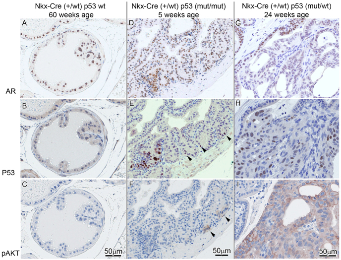
Fig. 2.
Lesion progression in Tp53R270H/+ Nkx3.1-Cre mouse prostate. (A–C) In Tp53 wild-type mice with Nkx3.1 haploinsufficiency, foci of atypia were seen in older mice (60 weeks) but are not associated with well-developed PIN. p53 (B) and pAKT (C) are negative. (D–F) Early pre-PIN atypia/PIN 1 lesion in a 5-week-old mouse with p53 mutation/stabilization as detected by IHC (bottom left and arrowheads in E) seems to precede pAKT (focally seen arrowheads in F) and AR expression in normal and atypical areas (D). (G–I) At 24 weeks, areas with p53 mutation/stabilization in PIN 3–4 are accompanied by pAKT expression (I) and decreased AR nuclear intensity and percentage (G).

