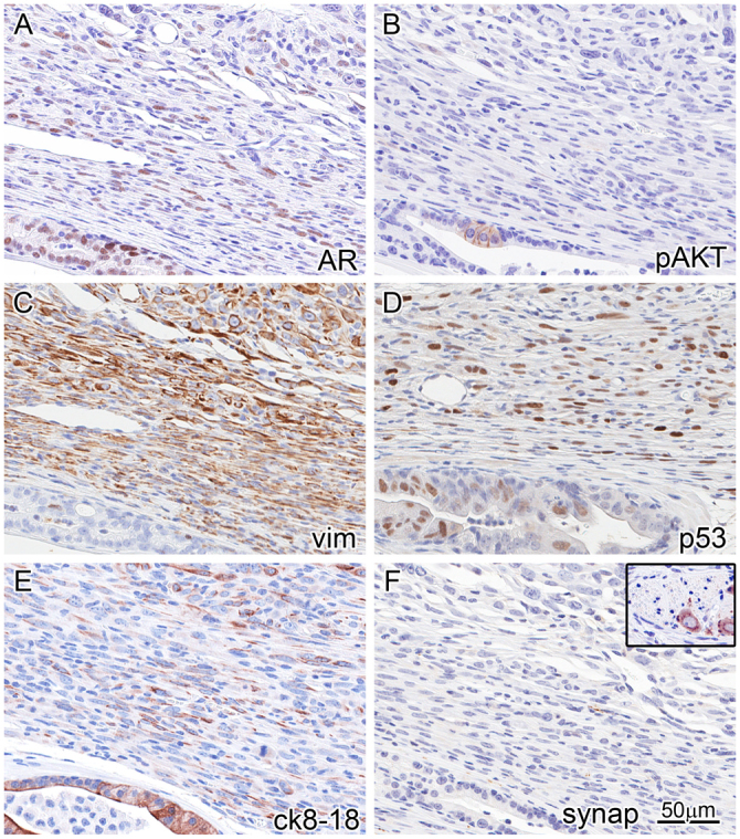
Fig. 4.

Immunophenotyping of invasive carcinoma reveals an EMT phenotype and AKT ‘de-addiction’. IHC (markers indicated lower right of each panel) on the same area (near-serial sections) of the periphery of an invasive tumor with an adjacent duct (bottom and lower left of each panel). This reveals strong AR expression in the duct, and weaker and lower percentage AR expression in the tumor cell nuclei (A). Focal pAKT is seen in the in situ atypia (B), corresponding to Tp53 stabilization/mutation (D). The tumor co-expresses vimentin (C) and luminal CK8/18 (E), and loses expression of pAKT (B). Although AKT activation is likely to be a driver of the PIN lesions, the invasive tumor seems to be independent of AKT (is ‘de-addicted’). The in situ areas are vimentin negative and cytokeratin positive (C,E). The tumor is negative for neuroendocrine differentiation as detected by a negative synaptophysin (F); inset is from the same tissue section showing a neural ganglion as internal positive control.
