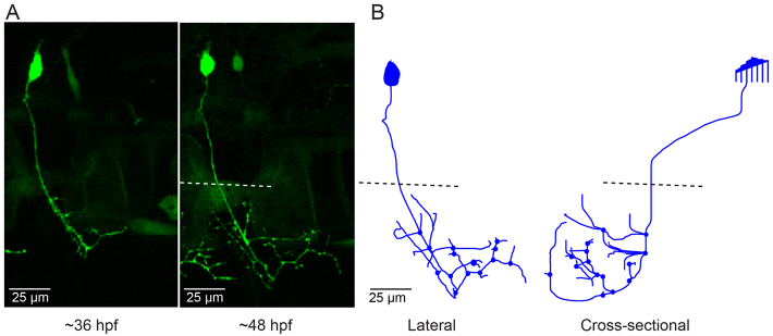
Fig. 4.
CaP neurons that express aoR3H tend to branch excessively at the distal end of the axon. (A) Projected confocal images obtained from an aoR3H-expressing CaP neuron at ∼36 and ∼48 hpf are shown. At ∼48 hpf, the distal axonal arbor is highly branched. (B) Projected Neurolucida trace of the ∼48 hpf image stack is shown from lateral (left) and cross-sectional (right) perspectives. See Fig. 1 legend for color code, etc.

