
Fig. 2.
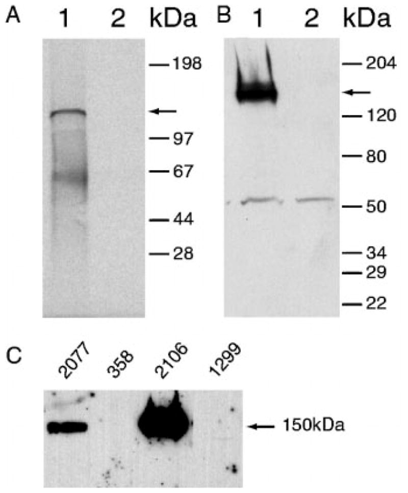
A, in vitro transcription and translation of α2δ-2. Lane 1, in vitro transcription and translation of α2δ-2 in expression vector pcDNA3.1. Lane 2, no DNA was added in the same reaction. The arrow indicates the expected 130 kDa product. B, Western blot analysis of transfection of NCI-H1299 cells with α2δ-2. Lane 1, transfection of NCI-H1299 cells with α2δ-2 in expression vector pcDNA3.1. Lane 2, transfection of NCI-H1299 cells with pcDNA3.1 vector alone. Affinity-purified anti-α2δ-2 peptide antibody was used to detect the protein product. The arrow indicates the expected protein product. Sizes of the prestained protein molecular weight markers are indicated on the right. C, 40 μg of protein from tumor cell lysates were loaded in each lane. Lane 1, NCI-H2O77 (adenocarcinoma); lane 2, NCI-H358 (adenocarcinoma); lane 3, NCI-H2106 (large cell neuroendocrine carcinoma); lane 4, NCI-H1299 (large cell carcinoma) cells.