
Figure 2.
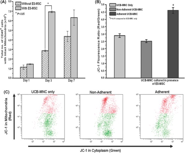
Effect of ES-MSC co-culture on the mitochondrial membrane potential of the UCB-MNC. (A) The mitochondrial membrane potential of UCB-MNC (combined fraction) over a time-course of 7 days, as determined by JC-1 staining. The number of cells with intact mitochondria was calculated from the red fluorescence-positive portion multiplied by the total number of cells (*P<0.05). (B) JC-1 red to green fluorescence ratio (red fluorescence mean/green fluorescence mean) in the non-adherent and adherent fractions of the UCB-MNC co-cultured with ES-MSC and non-co-cultured UCB-MNC over a 3-day culture period (*P<0.01). (C) Representative flow cytometer plots of the JC-1 staining performed on the non-adherent and adherent fractions of co-cultured (with ES-MSC) UCB-MNC compared with the non-co-cultured control. Data represent mean±SEM from three independent experiments.