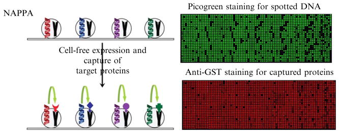
Figure 9.1.
On-array protein expression and capture. Left: Plasmid DNA is mixed with BSA, BS3 cross-linker, and the anti-GST capture antibody and arrayed on the aminosilane-coated glass slide. After blocking, cell-free expression mix is applied to the slide, and during a temperature-programmed incubation, the proteins are produced and bind to the capture antibody. Captured proteins can be detected by detecting the GST tag using a monoclonal anti-GST antibody, an HRP-labeled anti-mouse antibody, and Cy3-tyramide (TSA) HRP substrate. Right: Sample NAPPA images for PicoGreen staining of spotted DNA and anti-GST staining for captured proteins.

