
Figure 1. Electrical coupling and responses of SubC and PPN neurons.
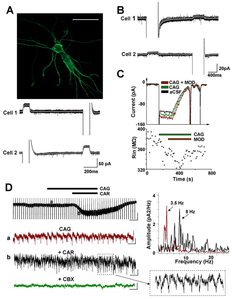
A. Electrical coupling in the SubC. Top: Two SubC neurons that were electrotonically coupled were imaged with neurobiotin (Cy2) immunofluorescence. The scale bar is 50μm. Bottom: Dual voltage clamp recordings (Holding Potential=-50mV) of the above neurons were conducted in the presence of TTX (1 μM). Hyperpolarizing pulses (-60 mV) injected into one cell induced an outward current in the neighboring cell, and vice versa. Gray line represents the average of 15 sweeps. The coupling ratio was calculated by dividing the outward current induced in one cell by the negative current injected into the neighboring cell. The coupling ratio of cell 1 to cell 2 was 8.13 ± 0.59 %, and of cell 2 to cell 1 was 6.25 ± 0.38 %. B. Whole-cell patch-clamp recordings from a pair of electrically coupled PPN neurons under voltage clamp. The same protocol as in A was applied, except that hyperpolarizing pulses were from -60 mV to -110 mV of 500 ms duration. The coupling ratio of cell 1 to cell 2 was 3.98 ± 0.55%, and of cell 2 to cell 1 was 4.08 ± 0.55%. C. An example of a PPN cell whose input resistance was decreased by fast synaptic blockers (CAG = 6-cyano-7-nitro-quinoxaline-2, 3-dione [CNQX] 10 μM, (±)-2-amino-5-phosphopentanoic acid [APV] 10 μM and gabazine 10 μM), then decreased further by the superfusion of modafinil (MOD, 150 μM). A 500 ms hyperpolarization step (from -60 mV to -105 mV) followed by a 1000 ms ramp (-105 mV to -35 mV) was applied in order to test the change of input resistance (Rin) and reversal potential of activated current. A higher current was required to compensate for the voltage change in the presence of modafinil, indicating a decrease in Rin (top record in red compared to control in black and in fast synaptic blockers in green). Rin changes during 12 min recording are shown on the bottom. D. Cholinergic modulation of spontaneous spikelets, indicative of electrical coupling. Left: In the presence of fast synaptic blockers (CAG), non-selective cholinergic receptor agonist, carbachol (CAR), induced 60 pA inward current in this PPN neuron with spontaneous oscillations (top, vertical bar 50 pA, horizontal bar 4 sec). Enlarged records from points (a) and (b) are shown below. Recording (a) showed spontaneous oscillations in the presence of CAG. Recording (b) demonstrated that CAR increased the frequency of oscillations. The box on the bottom right shows enlarged 1 sec record (b). Carbenoxolone, a gap junction blocker, completely blocked the oscillations (bottom). Blockade did not affect membrane potential. This suggested that the oscillations were modulated by electrical coupling. Scale bars for the enlarged records are vertical 10 pA and horizontal 500 ms. Top Right: Power spectrum histogram of the oscillations in (a) and (b). Each histogram was obtained from a 30 sec recording. The frequency of spontaneous oscillations was 3.5 Hz, which increased to 8 Hz following the application of CAR.