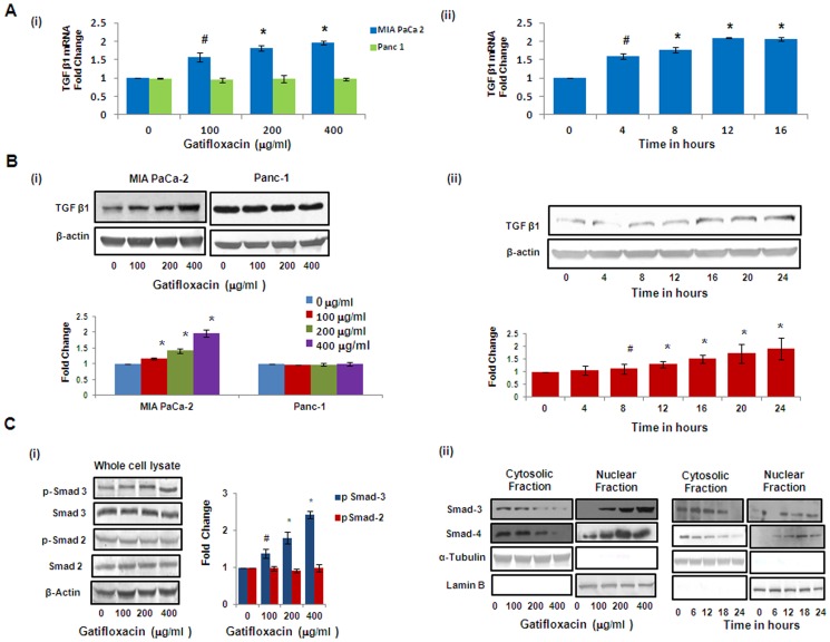
Figure 3. Gatifloxacin causes activation of TGF-β1 and Smad Complex in MIA PaCa-2.
(A) (i) Real Time PCR analysis of TGF-β1 expression in MIA PaCa-2 and Panc-1 cells treated with Gatifloxacin in a dose dependent manner, (ii) Real Time PCR analysis of TGF-β1 expression in MIA PaCa-2 cells treated with 400 µg/ml of Gatifloxacin in a time dependent manner. 18S rRNA was used to normalize the results. (B) Western blot analysis of TGF-β1 expression in MIA PaCa-2 and Panc-1 cells treated with Gatifloxacin in a dose dependent manner (i) western blot analysis of TGF-β1 expression in MIA PaCa-2 cells treated with 400 µg/ml of Gatifloxacin in a time dependent manner (ii). Data are representative of typical experiment repeated three times with similar results. Bar Graph represents the mean ± SEM. (C) (i) Effect of Gatifloxacin on receptor mediated Smads (pSmad-2 and pSmad-3) when assessed in whole cell lysate in MIA PaCa-2 cells. β-actin was used as a loading control. (ii) Translocation of Smad 3–4 complex from cytoplasm to nucleus under the effect of Gatifloxacin in a dose (0, 100, 200, 400 µg/ml) and time dependent manner (0, 6, 12, 18, 24 h) was assessed by western blotting. Nuclear and Cytoplasmic fractions were separated as described in the “Material and methods” section. Lamin B (Nuclear specific protein) and α-Tubulin (Cytoplasmic specific protein) were used as loading controls.

