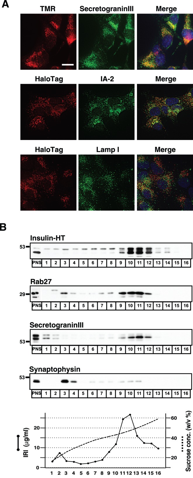
Figure 1. Insulin-HT targets to secretory granules in MIN6 cells.

A, MIN6 cells stably expressing insulin-HT (clone #67) were incubated with 5 µM HT-TMR probe for 30 minutes. The cells were then fixed and stained with anti-secretogranin III and Alexa488-conjugated anti-rabbit IgG antibodies (top panels). The stable cells were double-immunostained with anti-HaloTag and anti-IA-2 antibodies (middle panels) or anti-HaloTag and anti-lamp I antibodies (bottom panels). DAPI staining was performed for indentifying nucleus. Fluorescent images were captured by confocal microscopy. Bar, 10 µm. B, MIN6/insulin-HT cells were homogenized and the post-nuclear supernatants (PNS) were separated on a linear sucrose density gradient. Sixteen fractions were collected, and equal volumes from each were analyzed by immunoblotting with antibodies for HaloTag, rab27A/B, secretogranin III, or synaptophysin. Immunoreactive insulin (IRI) in a portion of each fraction was measured.
