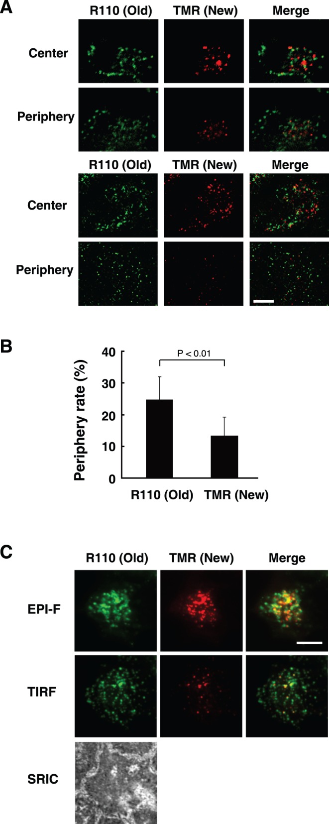
Figure 3. Secretory granules containing newly synthesized insulin are located at the interior of the cell.

A, MIN6/insulin-HT cells were incubated with 100 nM HT-R110 probe for 16 hours. The cells were then incubated in modified Krebs-Ringer buffer lacking dyes for 1 hour, and further incubated with 5 µM HT-TMR in buffer for 30 minutes. After removal of excess probe, the cells were fixed and fluorescent signals of R110 and TMR were observed by confocal microscopy in sequential z-axis planes. The periphery and the center images (upper panels) were from stage 2 and 9, respectively (full images were shown in Fig. S3). Another set of cell picture was shown in the lower panels. Bar, 10 µm. B, Intracellular localization of new or old insulin-HT was analyzed as in (A). Their periphery rates were quantified as described in Methods. Data are shown as the mean ± SEM of six independent experiments. C, Cells treated as in (A) were analyzed by total internal reflection fluorescence (TIRF) microscopy. Epifluorescence (non-confocal), TIRF, and surface reflection interference contrast (SRIC) images were simultaneously captured. Experiments were repeated three times with reproducible results. Bar, 10 µm.
