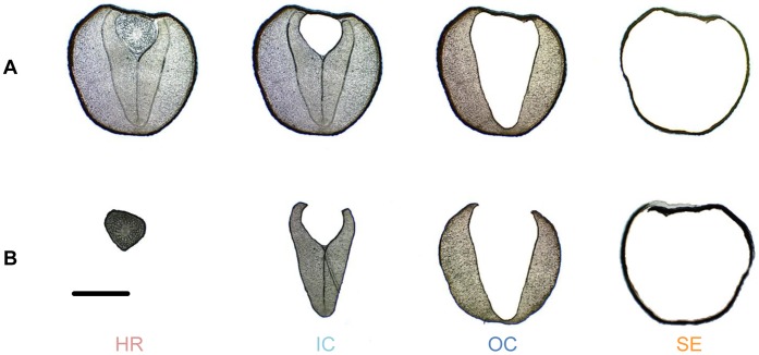
Figure 1. Work flow of laser microdissection of rapeseed.
(A) Progress of laser microdissection workflow applied to rapeseed. Hypocotyl and radicle (HR), inner cotyledon (IC), outer cotyledon (OC), seed coat and endosperm (SE) were successively dissected from rapeseed. (B) Micrographs of dissected tissues. Bar represents 1 mm.

