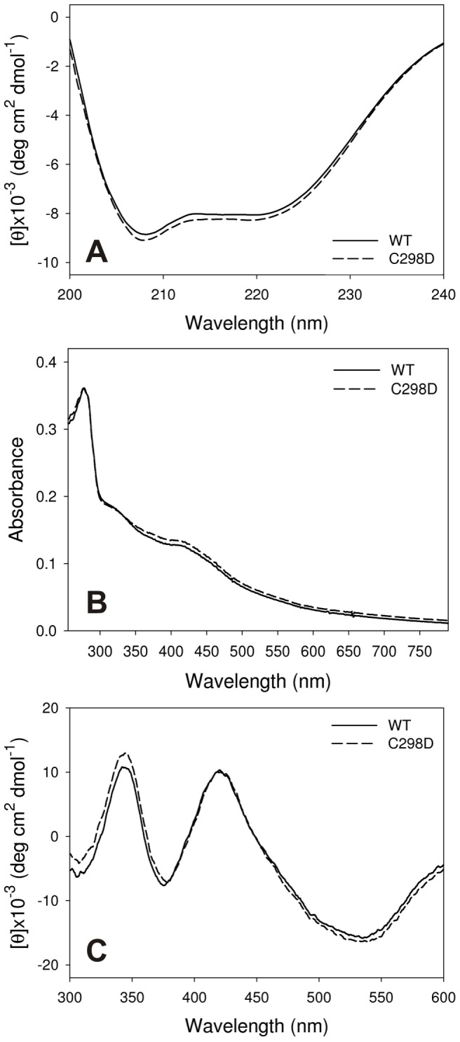
Figure 6. A) Far UV circular dichroism spectra of CaHydA WT (full line) and C298D (dashed line).

Spectra were acquired under aerobic conditions in 50 mM Tris·HCl, 200 mM KCl, pH 8.0. C298D mutation does not cause significant differences in secondary structure. B) UV-visible absorbance spectra of oxidised CaHydA WT (full line) and C298D (dashed line). Spectra were acquired under aerobic conditions in 50 mM Tris·HCl, 200 mM KCl, pH 8.0. C) Visible circular dichroism spectra of oxidised CaHydA WT (full line) and C298D (dashed line). A different behaviour is observed between 250–300 nm, whereas the visible region does not show any difference, demonstrating identical insertion of iron sulphur clusters.
