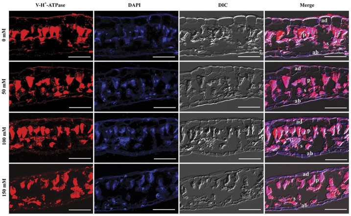
Figure 5. Immunolocalization of V-H+-ATPase subunit E in the leaves of B. papyrifera.
V-H+-ATPase subunit E was stained red with rabbit anti-VHA-E antibody and the nuclei were stained blue with DAPI. The merged images of VHA-E, nuclei and the DIC image are also presented. DIC, differential interference contrast; ad, adaxial epidermis; ab, abaxial epidermis; p, palisade tissue; s, spongy parenchyma. Scale bar = 50 μm.

