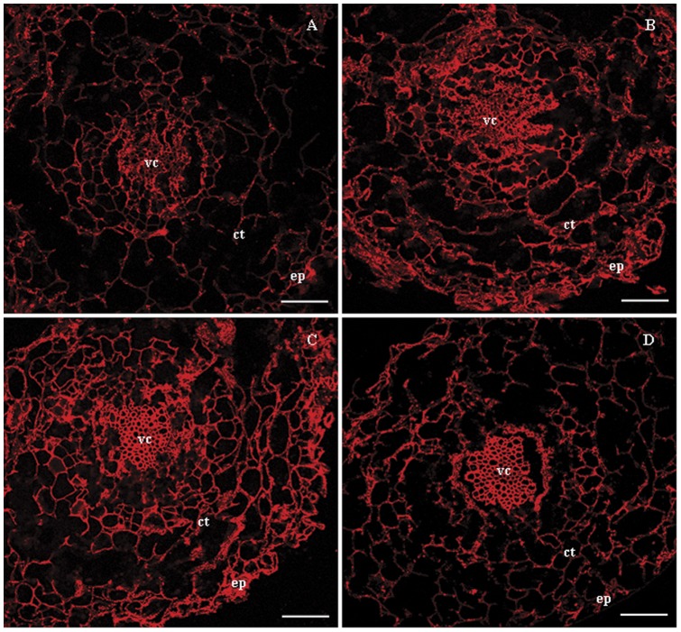
Figure 6. Distribution of V-H+-ATPase subunit E protein in root tissues of B. papyrifera grown under NaCl stress.
(A) Control, (B) 50 mM NaCl treated plants, (C) 100 mM NaCl treated plants and (D) 150 mM NaCl treated plants. Localization of VHA-E was examined by immunofluorescency using rabbit anti-VHA-E antibody. ep, epidermis; ct, cortex; vc, vascular cylinder. Scale bar = 50 μm.

