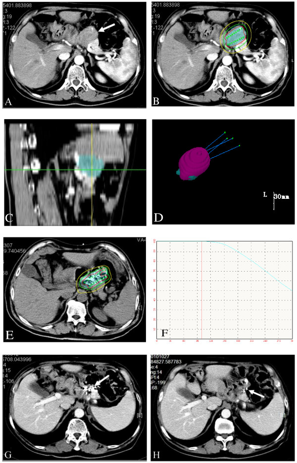
Figure 1.
TPS planning diagram and CT scans during follow-up with recurrence in the primary tumor bed after total gastrectomy. a). Before treatment, the tumor size was 35 mm in diameter; b). Isodose curves for treatment planning (“iso” 120 Gy, red line = 150%; green line =100%; yellow line = 50%); c). Sagittal images reconstructed by the TPS based on data from perioperation CT images: the skyblue area represent the PTV; d). Three-dimensional view of the TPS planning program: four puncture paths were designed; 99.0% of PTV (the skyblue area) was covered by 90% of isodose curves (the pink area); e). A typical CT slice showing the distribution of 125I seeds and isodose curves after seed implantation (red line = 180 Gy; green line =120 Gy; yellow line = 60 Gy); f). The D0 dose-volume histogram caculated by TPS (V100 = 96.2%, D90 = 117%); g). Two months after treatment, the recurrent tumor was completely eradicated, and 125I seeds gathered together; h). Six months after treatment, there was no progression in the treatment region, and some seeds migrated.

