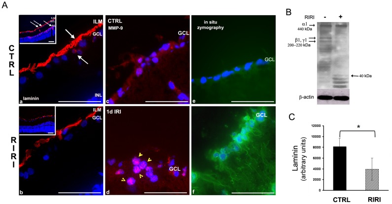
Figure 2. Laminin degradation is associated with increased MMP-9 expression and activity in the retina 1 day after RIRI.
A. Immunofluorescence with a pan-laminin antibody in retinal sections from control (CTRL), and ischemic (RIRI) eyes shows laminin expression (red) in the inner limiting membrane (ILM), ganglion cell layer (GCL), and inner nuclear layer (INL) (a, b, and lower magnification insets). White arrows in a, and corresponding lower magnification inset point toward sites of laminin degradation in the ECM of RGC, in the ILM, around the RGC cell body, and INL. Note thinning of laminin in the ILM, and loss of laminin expression in the RGC and INL after RIRI. (b, and lower magnification inset). Immunofluorescence with anti-MMP-9 antibodies shows increased expression of MMP-9 (red, yellow arrows; c, d), and in situ zymography demonstrates increased gelatinolytic activity (green) (e, f) in the ganglion cell layer (GCL) in ischemic eyes (n = 12). B. Western blotting of retinal extracts of control (−), and ischemic (+) rat eyes 1 day post-RIRI (n = 3). C. Densitometric analysis of bands corresponding to laminin β1 and γ1 chains in control and ischemic eyes normalized to β-actin. Error bars, SD. Student’s t test. *p<0.05. Scale bars: 100 µm.

