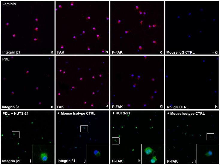
Figure 4. Laminin, or β1 integrin activating antibody, HUTS-21, promote integrin signaling in RGCs in vitro.
P5 RGCs were cultured on laminin (a–d), or PDL (e–l) for 48 hours. HUTS-21 (i, k) or isotype control (j, l) antibodies (1 µg/ml) were added to RGCs cultured on PDL 12 hours after plating. Immunohistochemistry was performed with β1 integrin (a, e, i, j), FAK (b, f), [pY397]-FAK (c, g, k, l), or IgG control antibodies (d, h) and analyzed by confocal microscopy. 100–200 cells per condition were analyzed. A representative experiment is shown. A total of N = 5 experiments were performed. Scale bars: 10 µm.

