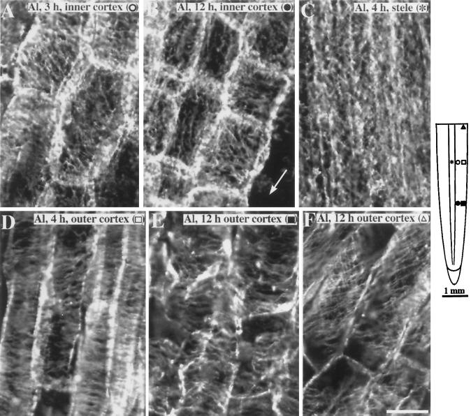
Figure 4.
Organization of cortical microtubules in maize primary roots after exposure to 50 μm Al. A, After 3 h of continuous exposure to Al, cells in the inner cortex 4 to 4.5 mm from the root tip showed random and obliquely oriented microtubules. B, After 12 h of continuous exposure to Al, reoriented microtubules occurred closer to the root tip. Cells in the inner cortex 2 mm from the tip showed random to longitudinally oriented microtubules. Arrow shows region where outer cortex has sloughed off. C, The stelar cells also exhibited random to longitudinal microtubules but occurred after 4 h of Al exposure. D, Four hours after Al exposure, outer cortical cells 4 mm from the root tip retained their net transverse microtubule orientation. E, Outer cortical cells 12 h after Al exposure also retained an overall transverse alignment of microtubules despite the distorted appearance of the cells. F, Cells in the maturation zone (about 6 mm from the tip) showed oblique microtubules that were similar to controls. The schematic diagram of the root indicates the regions where immunofluorescence images were obtained. Symbols in parentheses in A to F correspond to the symbols in the root diagram. Images are representative of at least five roots per time point. Bar in F = 25 μm.

