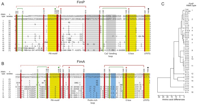
Figure 6. Sequence analyses of FimP and FimA among A. oris isolates.
A: Sequence alignment of FimP (n = 48) with fully conserved isopeptide bond triads (red), disulfide bonds (green), a conserved metal binding loop (grey) and pilin-, E-box- and LPLTG motifs in yellow. B: Sequence alignment of FimA (n = 14) with fully conserved isopeptide bond triads (red), disulfide bonds (green), a conserved proline-rich loop (blue) and pilin-, E-box- and LPLTG motifs in yellow. In addition, in A and B, polymorphic amino acid residues are shown (single letter codes). The top lines represent the consensus sequence and amino acid positions based on FimP and FimA respectively of reference strain T14V. C: Neighboring joining tree with sixteen allelic or sequence fimP types among A. oris isolates (n = 48) due to the single amino acid variations.

