
Figure 4. AT1b receptor deficiency had no effect on AngII-induced abdominal aortic dilation in vivo.
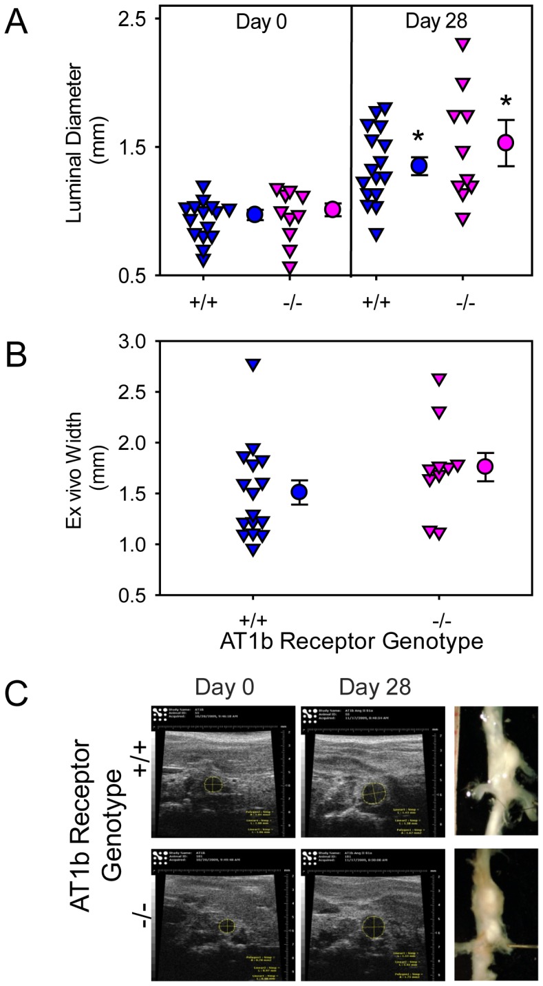
(A) Maximal luminal diameters of suprarenal aortas were measured in vivo by ultrasonography at baseline (Day 0) and on Day 28 during AngII infusion (N = 16: AT1b receptor +/+, and N = 10: AT1b receptor −/− mice). * denotes P<0.001 saline versus AngII within AT1b genotypes (two way repeated measures ANOVA). (B) Maximum width of supra-renal aortas was measured ex vivo (N = 16: AT1b receptor +/+, and N = 10: AT1b receptor −/− mice). Inverted triangles represent individual mice, circles represent means and error bars are SEM. (C) Examples of ultrasound images (Day 0 and Day 28) and ex vivo pictures (after termination) of suprarenal aortas, which represent aortic diameters nearest the mean of each group.