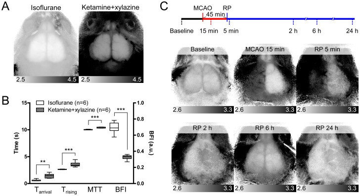
Figure 4. Detection of cerebral hemodynamic changes using CBF maps.
(A) Representative Trising maps for mice anesthetized with either 1.5% isoflurane or 0.1 mg/g ketamine and 0.01 mg/g xylazine. (B) Averaged CBF parameters of regions over left somatosensory cortices of six mice anesthetized with isoflurane and six mice anesthetized with ketamine and xylazine. Tarrival, Trising, and MTT parameters increased significantly, and BFI parameter was decreased significantly, in ketamine and xylazine group (t-test, **p<0.01; ***p<0.001). (C) Timeline of the transient MCAO protocol (upper diagram) and representative Trising maps (lower panels). Reperfusion (RP) was induced at 45 min after MCA occlusion. The six time points for imaging acquisition are indicated under the timeline.

