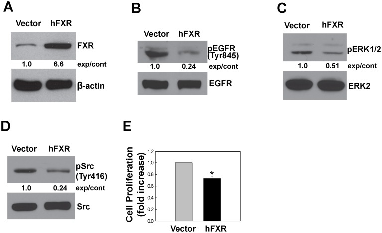
Figure 7. FXR overexpression inhibits phosphorylation of EGFR, ERK and Src, and attenuates cell proliferation.
A. Immunoblots showing FXR protein levels in HT-29 cells stably-transfected with empty vector alone or vector containing human FXR (hFXR). Phosphorylation of EGFR (Tyr845), ERK1/2, Src (Tyr416) as shown in panels B, C and D, respectively, was determined by immunoblotting with corresponding antibodies. Immunoblotting for β-actin, total EGFR, ERK2 or Src was used as a loading control. Numbers between immunoblots represent densitometry. Experimental/control (exp/cont) ratios were calculated after normalizing each test band to the respective control band. E. HT-29 cells stably-transfected with empty vector alone or hFXR were incubated for 2 days at 37°C. Cell proliferation was measured as described in Materials and Methods. Values represent mean ± SE from at least 3 separate experiments; *p<0.05 vs. control (Student’s t-test).

