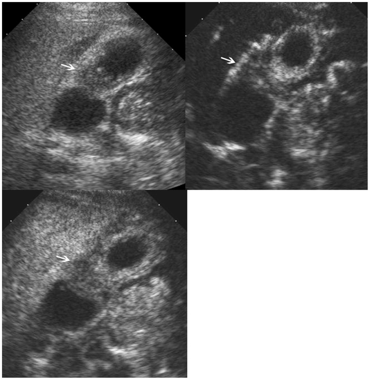
Figure 5. Adenomyomatosis in gallbladder.
Upper left, conventional ultrasound shows a slight hypoechoic mass (arrow) in the gallbladder; Upper right, the lesion (arrow) shows inhomogeneous hyper-enhancement during the arterial phase; Lower left, the lesion (arrow) shows hypo-enhancement during the venous phase.

