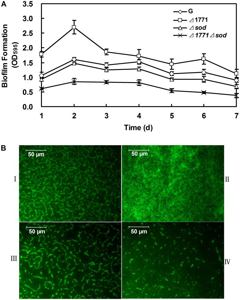
Figure 2. Quantification of the biofilms produced by different L. monocytogenes strains.
(A) Microtiter plate assay: Biofilm was measured each day for cells inoculated in TSB broth with 96-well microtiter plate for 7 days. The experiments were repeated three times and error bars indicate the standard deviations. The T-test was used to calculate the p-value between the wild-type and mutants. (B) Microscope assay with 0.1% FITC. I: wild-type L. monocytogenes 4b G; II: Δ1771; III: Δsod; VI: Δ1771Δsod. Cells from each strain for this method were incubated on glass slides for 3 days to form biofilms.

