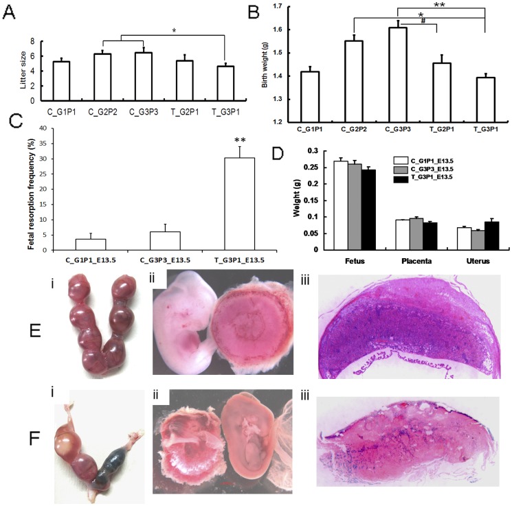
Figure 4. Impact of medical abortion on subsequent pregnancies.
Birth weight and litter sizes of F1 mice were recorded. Litters from mice that were subjected to repeated medical abortion showed decreased size. (A) The mean size of litters from T_G3P1 mice was significantly lower than those of C_G2P2 and C_G3P3 mice (T_G3P1 vs. C_G2P2 and C_G3P3, P<0.05). (B) The mean birth weight of litters from T_G3P1 was less than those of C_G2P2 and C_G3P3 mice (T_G3P1 vs. C_G2P2 and C_G3P3, P<0.05 and P<0.01, respectively). The mean birth weight of T_G2P1 litters was significantly lower than that of C_G3P3 litters (P<0.05). (C) The pregnancy status of T_G3P1 mice was examined at 13.5 days of gestation. T_G3P1 mice had significantly increased fetal resorption frequencies compared with C_G1P1 and C_G3P3 mice (P<0.01). (D) The weight of the placenta, fetus, and uterus was also measured, but no statistically significant differences were observed between the control and treatment groups. (Ei and Fi) Fetal resorption was compared between the T_G3P1_E13.5 (Fi) and control group (Ei) BALB/c mice after 13.5 days of gestation. (Fii) Compared to the control groups (Eii), some placentas were abnormal, and necrosis and fetal death were observed in pregnant T_G3P1 mice. (Fiii) The placentas appeared to be small labyrinths, with degenerated structures observed in T_G3P1 mice, but not in control groups (Eiii).

