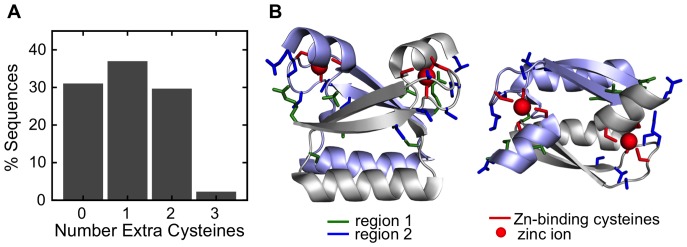
Figure 4. Cysteine-rich regions in the E7C domain.
(A) Distribution of the number of cysteines in the cysteine-rich regions of individual E7C domains. (B) Ribbon representation of the average NMR structure of HPV45 E7C (PDB ID: 2F8B), with cysteine-rich positions corresponding to regions 1 (green) and 2 (blue) in stick representation (see text). Note that for many cysteine-rich positions the corresponding HPV45 E7C residue is not a cysteine. Zinc atoms are represented as red spheres. Protein representations were generated using Pymol (http://www.pymol.org).

