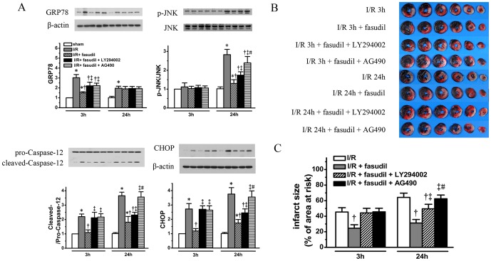
Figure 9. To roles of PI3K/Akt and JAK2/STAT3 signaling in ER stress modulated by fasudil were also evaluated.
Using the same heart tissue obtained as described as in Fig. 5 and Fig. 6, ER stress related apoptosis protein markers, including GRP78, phospho-JNK, cleaved Caspase-12 and CHOP were quantified (panel A). IS were measured for I/R rats receiving fasudil with either LY294002 or AG490 at both 3 and 24 hours (n = 8 at each time point per group), the results were compared with I/R group and I/R with fasudil group (using the same data from figure 2). Representative heart slices with TTC staining are shown in panel B with quantification shown in bar graph (Panel C). All data shown are expressed as mean±SD. * denotes P<0.05 vs. sham group at the same time point; †, P<0.05 vs. I/R group at the same time point; ‡, P<0.05 vs. I/R+fasudil group at the same time point and #, P<0.05, vs. I/R+fasudil+LY294002 group at 24 hours of reperfusion.

