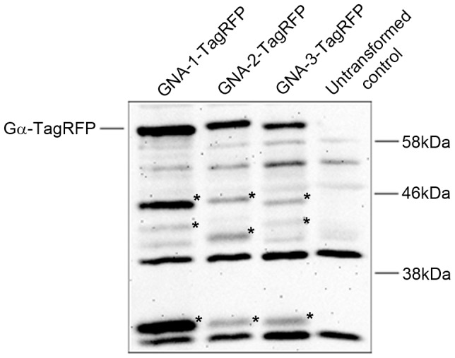
Figure 7. Western blot detection of Gα-TagRFP fusion proteins.

Samples containing 50 µg of protein from conidial extracts were subjected to western blot analysis with a RFP primary antiserum as described in the Materials and Methods. Strains are Δgna-1, gna-1-TagRFP (2.1), Δgna-2, gna-2-TagRFP (5.1), Δgna-3, gna-3-TagRFP (12.1) and wild type (untransformed control; 74-OR23-IVA). TagRFP is 27 kD, while the predicted size of the three TagRFP fusion proteins is 68 kD. Potential degradation products for each RFP fusion are noted with asterisks.
