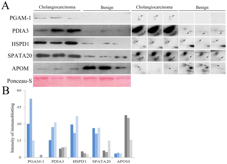
Figure 2. Validation of the 2-DE results by Western blotting.
(A) Left panel: Western blotting analysis of aliquots of pooled bile proteins from patients with CC or cholangitis. Right panel: the corresponding spots, with the same molecular weights, in the 2-DE gels. The Ponceau-S-stained blot (below) was used the internal control. (B) Quantification of protein expression in pooled bile proteins. The bars represent the signal intensity of the immunobands and were consistent with the 2-DE results.

