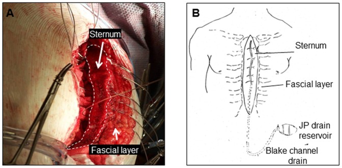
Figure 1. Fluid and cell collection from sternal wound environment.
A, image of the sternal wound before closure where the Blake drain is yet to be placed; B, after closure of the sternum, the surgeon placed a Blake drain over the sternum, and then closed the wound in layers. The Blake drain was connected to a heparinized J-VAC bulb suction reservoir for wound fluid collection.

