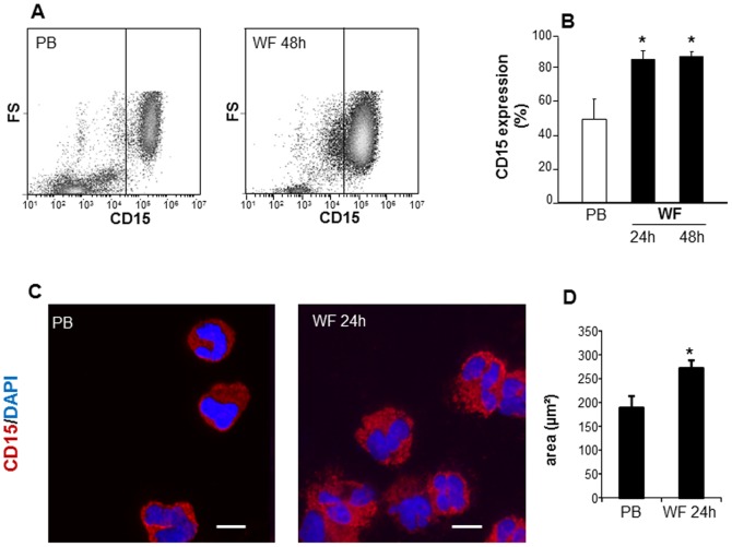
Figure 3. PMN in wound fluid are CD15 positive.
PB and WF leukocytes were isolated and stained with FITC-conjugated anti-human CD15 followed by A–B, flow cytometry analysis or C–D, cytospin. A, representative flowcytometry plots showing increased CD15 positive cells in WF at 48 h post wounding as compared to peripheral blood at baseline (PB). B, CD15 expression analysis using flow cytometry. Data presented as mean±SD, n = 4. *p<0.05 compared to PB. C, Representative images of cytospun cells derived from WF or PB. Cells were immunostained with CD15 (red) and DAPI (nucleus, blue). Scale bar = 10 µm; D, size analysis of cytospun cells derived from WF and PB. Data are mean ± SD; p<0.05 compared to PB.

