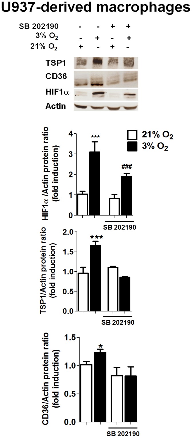
Figure 2. Hypoxia induces TSP-1 and CD36 expression and HIF-1α stabilization through activation of p38-MAPK.
U937 cells were maintained under normoxia or hypoxia in the presence or absence of SB 202190 (a p38-MAPK inhibitor, 10 µM, 24 h) and levels of proteins were determined by Western blot. Graphs show quantification of HIF-1α, TSP-1 and CD36 by densitometry. In hypoxia, cells treated with SB 202190 exhibited significantly lower protein expression of HIF-1α, TSP-1 and CD36 than cells treated with vehicle. In all cases bars represent mean± SEM (n>3). Comparisons between groups were performed using ANOVA followed by a Newman Keuls test. *P<0.05 and ***P<0.001 with respect to all groups in the same graph and ###P<0.001 vs. bars in normoxia.

