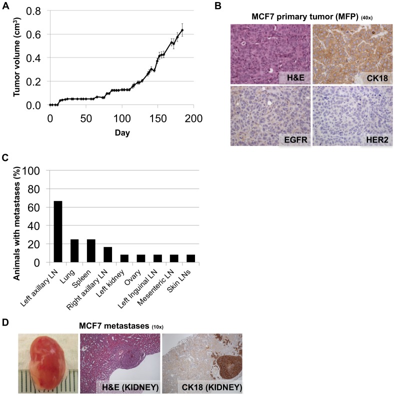
Figure 3. NSG mice occasionally develop macro-metastases when MCF7 cells are injected orthotopically into the mammary fat pad.
A. Volumes of tumors in mammary fat pads of NSG mice injected with MCF7 cells. Each data point is the mean value (+/− s.e.m) of 12 primary tumors. B. Micrographs of haematoxylin and eosin (H&E), CK18, EGFR and Her2 IHC staining of harvested MCF7 primary tumor tissue. MCF7 primary tumors are CK18 positive, EGFR negative, Her2 negative. C. Quantification of the percentage of mice bearing macro-metastasis in each organ observed at the time of necropsy (184 days post injection). Macro-metastases were observed in the left axillary lymph node (67% of mice), lung (25% of mice), and spleen (25% of mice), as well as sporadically in other organs. LN = lymph node. D. A photograph of an MCF7 kidney metastasis is shown, along with micrographs of H&E and CK18 IHC staining of harvested tissue.

