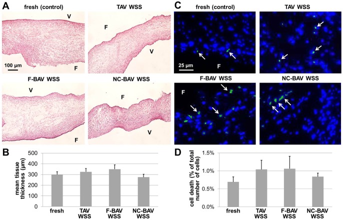
Figure 4. Effects of TAV and BAV WSS on tissue structure and cell viability.
H&E stain (A), mean tissue thickness measurements (B), TUNEL assay (C) and quantitative TUNEL results (D) in porcine aortic valve leaflets subjected to TAV and BAV WSS (F: fibrosa; V: ventricularis, green: apoptotic cells, blue: cell nuclei).

