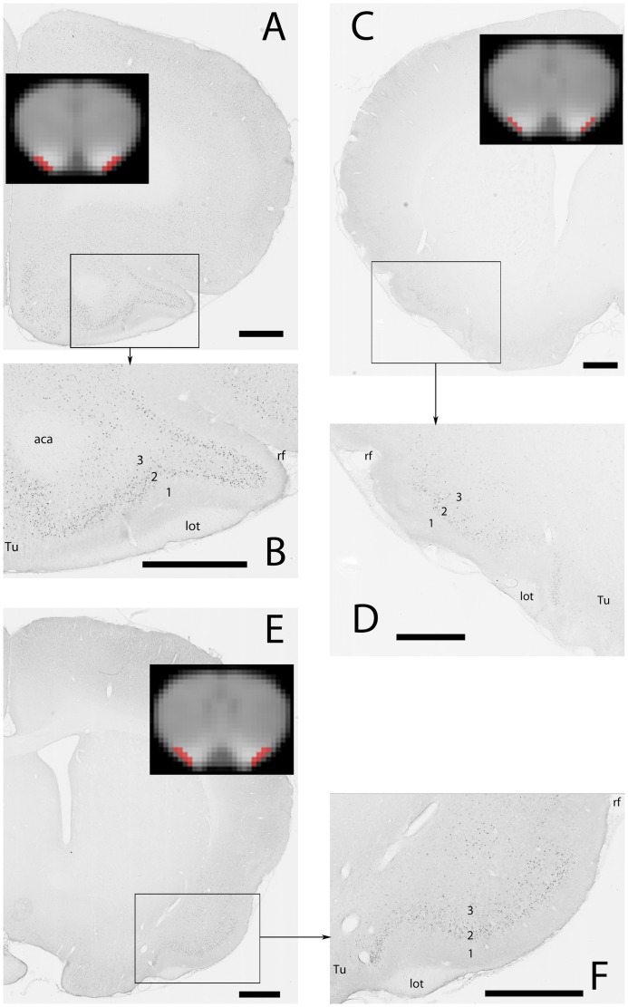
Figure 3. MEMRI contrast enhancement and c-Fos immunodetection in the anterior piriform cortex.
A, C & E: photomicrographs of the right (C) and left (A, E) sides of the brain. MEMRI images of the corresponding slices are shown in inserts with the ROI encompassing the piriform cortex in red. B, D & F: enlargements of the regions delimited by the black rectangles in A, C & E, respectively. A, B: anterior region of the anterior piriform cortex; stimulation: chocolate flavored cereals C, D: medial region of the anterior piriform cortex; stimulation: empty container E, F: posterior region of the anterior piriform cortex; stimulation: fox feces Abbreviations (1), (2), (3): layers 1, 2 and 3 of the piriform cortex, (aca) anterior part of the anterior commissure, (lot) lateral olfactory tract, (rf) rhinal fissure, (Tu) olfactory tubercle. Scale bars (only for Fos images): 1 mm.

