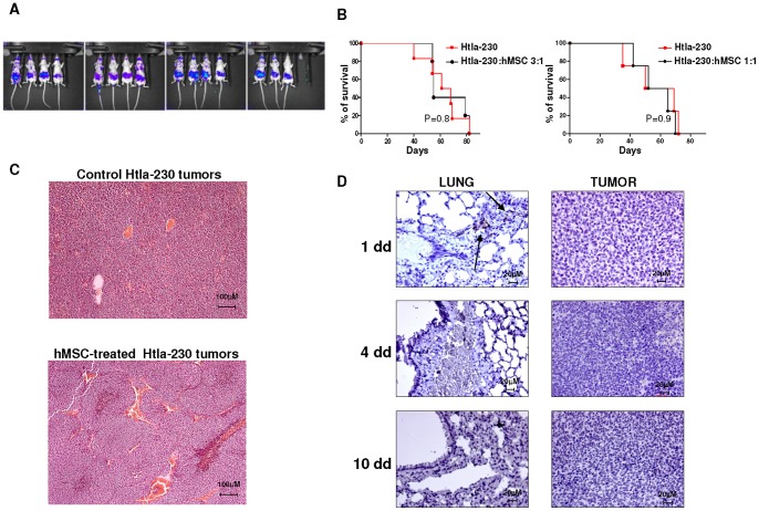
Figure 1. Survival curves and immunohistochemical analysis of human Htla-230 neuroblastoma bearing mice treated with human mesenchymal stem cells.
Panel A. Athymic mice (Nude-nu) were i.v. injected with ffLUC-Htla-230 (3×10⁶ cell/mouse; n = 15). Tumor establishment occurred 14 days after tumor cell inoculum as assessed by bioluminescence imaging. Panel B. Athymic mice (Nude-nu) were injected in the tail vein with Htla-230 (3×10⁶ cell/mouse) and treated i.v. with hMSCs (1×10⁶ cells/mouse, n = 6 or 3×10⁶ cells/mouse, n = 5) or saline solution (n = 6) 14 days after tumor cell inoculum. Survival curves were constructed by using the Kaplan–Meier method. Statistical analysis of different treatment groups was performed by Peto's log-rank test. Panel C. Representative hematoxylin-eosin-staining of tumors surgically removed from control and hMSC-treated Htla-230-bearing mice is shown. Original magnification 10x. Panel D. Representative immunohistochemical CD90 staining of lungs and tumors surgically removed from hMSC-treated Htla-230 bearing mice at different times after hMSC i.v. inoculum. Arrows indicate hMSCs positive for CD90. Original magnification 40x.

