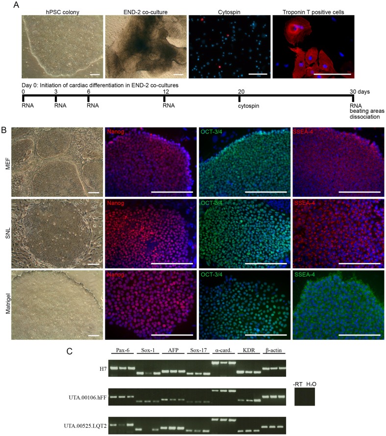
Figure 1. hPSCs were cultured with three different culture methods.
A) Schematic presentation of cardiac differentiation in END-2 co-culture and the experimental design. hPSC = human pluripotent stem cell, END-2 = mouse visceral endodermal-like cells. Scale bars, 200 μm. B) All five hPSC lines cultured on MEF and SNL feeder cell layers in conventional stem cell culture medium and on Matrigel in mTeSR1 medium at least for 14 passages formed undifferentiated colonies, which expressed pluripotency markers Nanog, OCT-3/4 and SSEA-4. Representative images of UTA.00112.hFF (phase contrast microscope images) and H7 (immunofluorescence images) cell lines are presented. Scale bars, 200 μm. C) H7, UTA.00106.hFF and UTA.00525.LQT2 cell lines cultured in all three culture conditions (in figure: first band MEF, second band SNL, third band Matrigel) formed embryoid bodies (EBs) expressing markers from all germ layers: ectoderm (PAX-6 and SOX-1), endoderm (AFP and SOX-17) and mesoderm (α-cardiac actin and KDR).

