Figure 1.

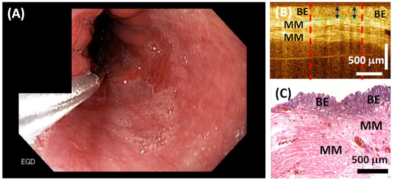
(A) Representative endoscopic image of the GEJ before RFA treatment. (B) Representative cross-sectional OCT image and (C) corresponding histology illustrating the BE epithelium thickness measurement.

(A) Representative endoscopic image of the GEJ before RFA treatment. (B) Representative cross-sectional OCT image and (C) corresponding histology illustrating the BE epithelium thickness measurement.