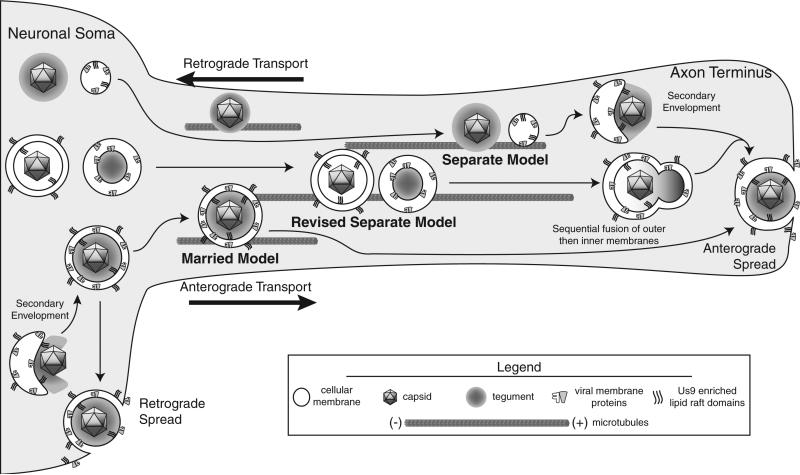
Figure 1.
Schematic of HSV anterograde transport models. A schematic visualization of the Married, Separate, and Revised Separate Models of anterograde transport. Particles are diagramed at three stages: cell body assembly/sorting, axonal transport, and egress. Distribution of tegument is not accounted for by the Revised Separate Model and therefore not presented

