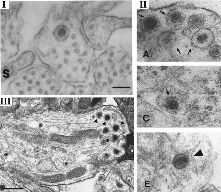
Figure 2.
Comparison of EM micrographs of putative naked capsids, dense-core vesicles, and enveloped virions in axons. (I) Figure 3, from Penfold et al. [35]—“unenveloped nucleocapsid in close proximity to microtubules” Copyright (1994) National Academy of Sciences, USA, reproduced with permission. (II) Figure 4, Panels A,C,E, from Holland et al. [59]—“TEM of axons in the . . . noninfected (A to C) model at 24h postinfection; (E) Viral nucleocapsids in the infected model” Copyright (1999) American Society for Microbiology, reproduced with permission. (III) Figure 1, Panel D, from Negatsch et al. [34]—“Black triangles indicate enveloped virions; . . . Neurovesicles are marked by lozenges.” Copyright (2010) American Society for Microbiology, reproduced with permission. Note: quotations indicate excerpt from authors’ original figure legend. The ability to discern between enveloped and naked capsids in axons through EM has been confounded by the presence of dense-core vesicles. Note the similarity between the naked capsid described in Penfold et al. [35] and the dense-core vesicles in Holland et al. [59]. Note the absence of naked capsids and the clear distinction between enveloped virions and dense core vesicles made possible by high-resolution EM in Negatsch et al. 2010 [34]

