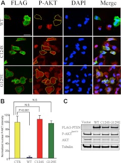
Fig. 4.
PTEN suppresses nuclear AKT, depending on its lipid phosphatase activity. A, BT-549 cells were transfected for 24 h with various FLAG-tagged PTEN constructs, as noted. Cells were fixed and stained with FLAG (green) and P-AKTSer473 (red) antibodies and were visualized by confocal microscopy. Dashed lines demarcate BT-549 cells expressing FLAG-PTEN. DAPI, 4′,6′-diamino-2-phenylindole. Scale bar, 50 μm. B, Average intensities of nuclear P-AKT of transfected cells were normalized to that of nontransfected cells in the same field. For each group, 20 positively stained cells were quantified for immunofluorescent intensities. Average intensities of nuclear P-AKTSer473 were plotted on a graph, and the statistical analysis was performed using one-way ANOVA. The error bars represent the sd. N.S., Not significant. C, BT-549 cells were transiently transfected with FLAG-tagged PTEN constructs, as noted. Western blots showed that only wild-type PTEN suppresses P-AKTSer473.

