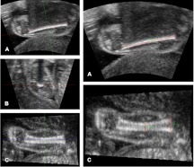
Fig. 1.
Femur measurement technique using 3D ultrasound and the MITK software (near field at the bottom of the image). Before measurement, the orthogonal orientation axes are aligned with the longitudinal axis of the femur in planes A and C and their intersection positioned at the midshaft (left, A, B, C). The coronal A plane is then used for FL measurement (right A), and the sagittal C plane is used for PMD and MSD measurement (right C).

