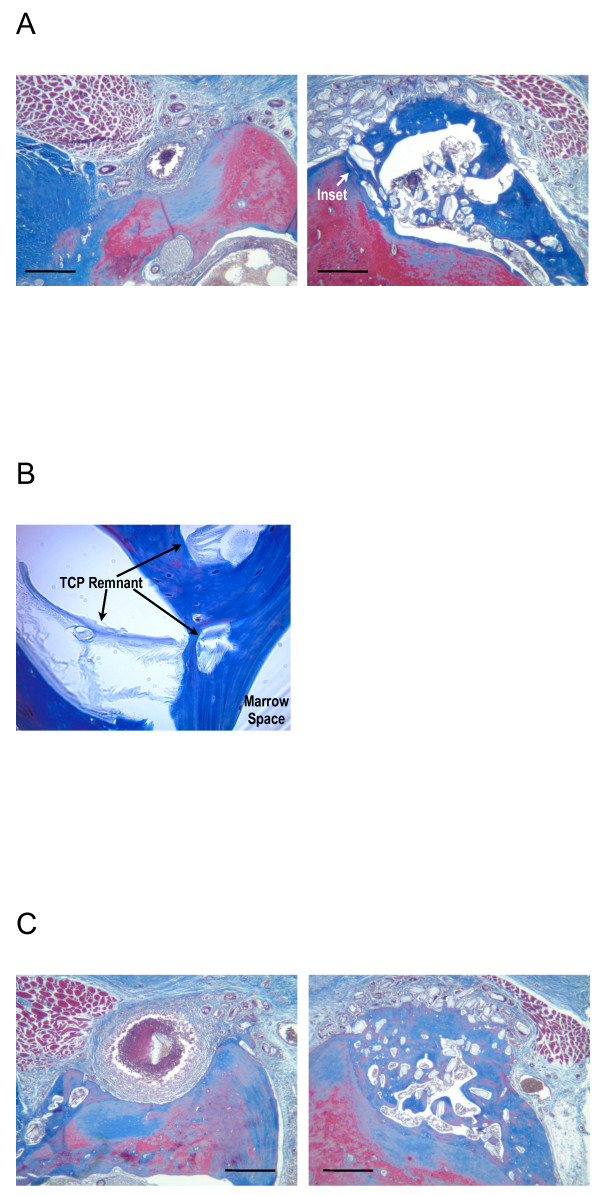
Figure 10.
Masson’s Trichrome stain, 12 wk healing.A: Left - TCP loaded and lyophilized with 50 μg/ml BMP-2. Right - B-247 treatment of TCP loaded and lyophilized with 50 μg/ml rhBMP-2 plus 100 μg/ml of pln.247 plasmid. Inset arrow designates region of panel B. B: 400x magnification of region designated in panel A between crest of ridge (towards left) and marrow space. Arrows designate remnant TCP particles which remain partially integrated with new bone (blue) after 12 weeks. C: Left - TCP loaded and lyophilized with 50 μg/ml BMP-2. Right - B-247 treatment of TCP loaded and lyophilized with 50 μg/ml rhBMP-2 plus 100 μg/ml of pln.247 plasmid. Pairs of images are from the same animal. Bar = 0.5 mm.

