
Figure 1.
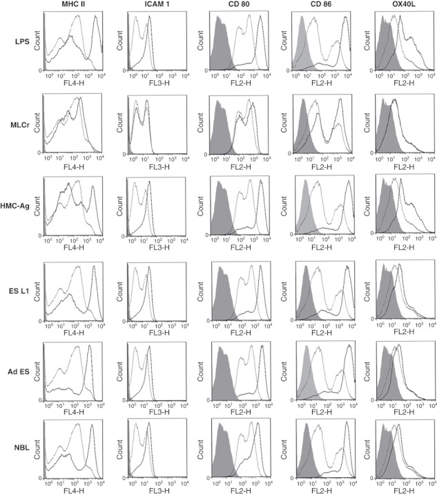
Expression of surface markers on mouse BMDCs pulsed with LPS and Trichinella spiralis antigens for 48 hrs (full dark line) as compared with untreated cells (dotted line). Isotype controls are shown with light grey shading. Results are representative of five experiments, each of which gave similar results.