
Figure 5.
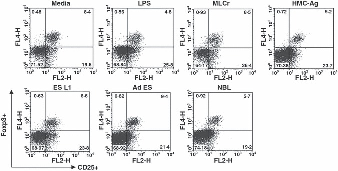
Foxp3+ cells within the CD4+ population of T cells isolated from mesenteric lymph nodes of C57BL/6 mice cultured with BMDCs (non-stimulated and stimulated with LPS or Trichinella spiralis antigens) for 7 days in presence of recombinant mouse IL-2 and anti-CD3 antibody. Results are representative of four experiments, each of which gave similar results.