Fig 2.
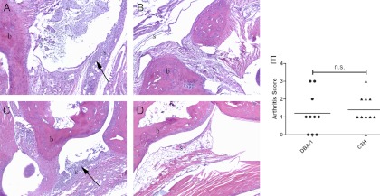
DBA/1 and C3H/HeJ mice develop similar histologic evidence of arthritis in tibiotarsal joints. DBA/1 and C3H/HeJ (n = 10 each) tibiotarsal joints were collected at 42 days after B. burgdorferi infection or sham infection and then decalcified and H&E stained. (A to D) Representative images at a magnification of ×10 are shown for B. burgdorferi-infected (A) or sham-infected (B) DBA/1 mice and for B. burgdorferi-infected (C) or sham-infected (D) C3H/HeJ mice. b, bone; s, synovium. Proliferative synovitis with leukocyte infiltrates is indicated by arrows. (E) Histologic scoring of arthritis severity was not different for the two strains. n.s., not significant (P = 0.66).

