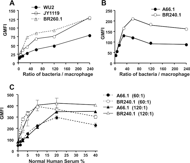
Fig 2.
Effect of PspA expression on the phagocytosis of pneumococci by macrophages. The phagocytosis of WU2 (A) and A66.1 (B) was examined at different ratios of bacterial CFU to macrophage in the presence of 10% normal human serum. FITC-labeled pneumococci were incubated with mouse macrophage cell line J774A.1 with a ratio of bacterial CFU to macrophage ranging from 240:1 to 4:1. Trypan blue solution was used to quench fluorescence from pneumococci adherent to the cell surface, thus only fluorescence from ingested pneumococci was measured. Each of these studies was repeated at least three times with similar results. Results of representative experiments are presented. To be able to evaluate these data statistically, the data points for each replicate were ranked. The ranked data points for each replicate for the portions of the curves that discriminated most strongly among the strains (bacterium/macrophage ratios of 30:240 for panel A and 60:24 for panel B) were pooled and analyzed by the Mann-Whitney two-sample rank test. In both experiments the differences between the wild-type and mutant strains were significant at P < 0.003. (C) Effect of the amount of available complement on pneumococcal phagocytosis of A66.1 and BR240.1. FITC-labeled bacteria were incubated with the indicated concentrations of normal human serum and then incubated with macrophage with ratios of bacterial CFU to macrophage of 60:1 and 120:1. Standard errors are indicated. The absence of visible standard error bars indicates that they were closer together than the width of the respective symbols. All flow cytometry data were based on ≥20,000 gated events.

