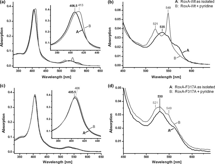
Fig 4.
UV-vis spectra of WT-RoxA and RoxA-F317A with heme ligands. (a and b) Spectra of recombinant WT-RoxA, as isolated (2 μM) and after addition of pyridine (5 mM; 1 h at room temperature), at the endpoint of the reaction. The Soret-band region is enlarged in the inset of panel a; panel b shows the Q-band region from panel a. WT-RoxA rapidly reacted with pyridine to give characteristic spectral changes as formerly reported (18), in accordance with a partial reduction of one heme center. (c and d) Spectra of recombinant RoxA-Phe317Ala, as isolated (2 μM) and after addition of pyridine (5 mM; 1 h at room temperature). In panel c, the Soret-band region is enlarged in the inset; panel d shows the Q-band region from panel c. Only minor changes appeared in the presence of pyridine and did not proceed through a prolonged incubation time. Similar weak effects on the UV-vis spectrum of RoxA-Phe317Ala were obtained by addition of other external heme ligands, such as imidazole (not shown). The characteristic changes in the WT-RoxA spectrum caused by imidazole have been reported previously (see Fig. 3a of reference 18).

