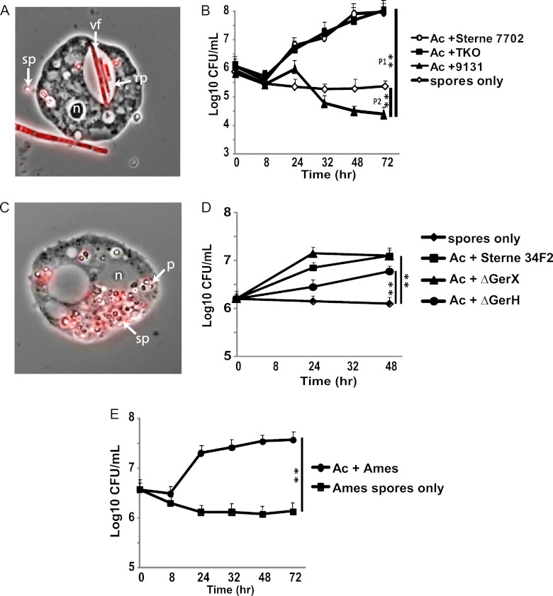
Fig 1.
Intracellular germination and growth of B. anthracis in coculture with A. castellanii. (A) Micrograph (magnification, ×100) of A. castellanii infected with RFP-expressing Sterne 7702 spores (red) after 6 h at 37°C. rp, replicative phagosome containing vegetative B. anthracis; sp, spores; vf, vegetative form; n, nucleus. (B) Growth of B. anthracis Sterne 7702, TKO, and 9131 with or without (spores only) A. castellanii (Ac) at the indicated times at 37°C. Data are the means ± standard errors of the means (n = 4 to 6; performed in triplicate). Statistical differences were determined by Student's t test comparing the indicated strain to the Sterne strain cultured in the absence of amoebas at 72 h (**; P1 = 0.000285; P2 = 0.00105). (C) Phase-contrast micrograph (magnification, ×100) after 6 h of culture at 37°C of A. castellanii with fluorescently labeled strain 9131(red spores). p, phagosome containing B. anthracis spores; sp, spores; n, nucleus. (D) Similar to the experiment described for panel B but with B. anthracis Sterne 34F2 as the parental strain control for mutants with either GerX or GerH eliminated. Data are the means ± standard errors of the means (n = 4). Statistical differences were determined by a Student's t test (**, P < 0.001). (E) Similar to the experiment described for panel B but with the B. anthracis Ames strain with or without A. castellanii. Data are the means ± standard errors of the means (n = 4). Statistical differences were determined by a Student's t test (**, P = 0.000356).

