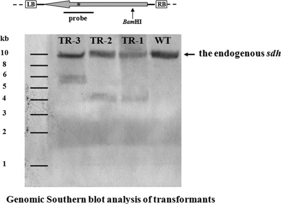
Fig 3.

Detection of wild-type and transgenic mutant copies of sdhB in three randomly selected G. lucidum transformants. (Top) Schematic representation of the restriction enzyme-probe combination used to detect the transgenic mutated allele of sdhB. White boxes are the left and right border regions of T-DNA, respectively. The large gray arrow represents the carboxin resistance cassette cbxr. An asterisk (*) indicates the position of single nucleotide mutation responsible for carboxin resistance in sdhB. The position of the single BamHI site within the T-DNA is indicated by a black arrow. (Bottom) Random integration of T-DNA copies into the G. lucidum genome. WT, G. lucidum wild-type strain; TR-1 to TR-3, independent transformants obtained after ATMT using plasmid pJW. The genomic copy of the sdhB is indicated by arrow on the right.
